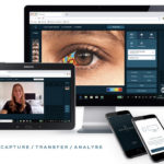
Technology from Advanced Ophthalmic Systems (AOS) allows Dr. Lang and his colleagues to deliver efficient, high-quality exams.
Sponsored Content
By Jacob R. Lang, OD, FAAO
August 17, 2022
Impressing upon patients the importance of treatment for dry eye and other conditions can be challenging. With patients exposed to so much health information online and elsewhere, it’s sometimes hard to get them to remember and absorb the information you present.
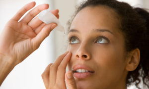
Fortunately, our practice invested in transformative digital eyecare technology from Advanced Ophthalmic Systems (AOS). It allows us to quickly and easily take photos of patients’ eyes. These images make such a strong impression on patients that they almost always sign on for the full treatment plan we prescribed.
The ability for the technology to quickly capture that kind of information makes all the difference during a busy time like the back-to-school season, when we strive to keep patient flow moving while delivering advanced eyecare.
High-Quality Images Efficiently Captured
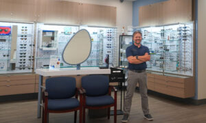
I see around 35 patients per day on average. During back-to-school season, that number hovers closer to 40, or a little more. We are typically 100 percent booked late July through the beginning of September with children returning to school needing care, as well as young adults who want to get in for an eye exam before heading back to college out of town. Our practice includes nine optometrists and eight ophthalmologists, so saying we’re usually completely booked this time of year is no small thing.
The high volume of patients we see in late summer/early fall doesn’t mean we are willing to sacrifice exam quality. We want to carefully screen our patients for both sight-threatening and quality-of-life threatening conditions, like dry eye. That’s where the technology we invested in from AOS a few years ago comes in.
Show Both the Need & Impact of Treatment
When you can show in-depth, clear photos of a patient’s eyes that first show exactly what’s happening in a patient’s meibomian glands, for example, and then show after treatment, how that same aspect of the patient’s eyes look, you have an invaluable patient compliance tool.
The best part is I don’t find that the capturing of these high-quality photos negatively impacts our patient flow. The technology is simple to use, so that our support staff could easily capture the photos, leaving the doctor to interpret the results. I currently take the photos myself. However, the staff can easily capture photos with the technology, which my colleagues and I could then interpret and provide recommendations on afterwards. This possibility offers us a chance to extend the opportunity to efficiently deliver a high level of care in a fast-paced environment.
We get a lot of “wows” from patients, who have been told for years that they have dry eye, but before seeing these photos, never understood what that really meant, why the condition is negatively impacting their vision and comfort, and why and how treatment will help.
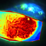
AOS fluorescein analysis of corneal limbal stem cell deficiency helps Dr. Lang’s patient understand the diagnosis better. The image shows the fluorescein pattern in 2D, highlighting the effect the condition has on the corneal surface.
The technology is able to provide objective analysis that shows output in tenths, so patients can see incremental progress, even if it’s small, which validates that they are on the right treatment plan. This is helpful in dry eye especially, with in-office treatments that may take 2-3 visits to show major progress.
Create More Efficient Follow-Up Appointments
The photos taken with AOS technology lay a foundation of knowledge so that we don’t have to start from the beginning in patient education at the next appointment. The patient, after seeing these images, and having the doctor explain what they are seeing, usually fully understands what is going to happen at their next appointment(s).
The time we take on the front end to educate patients, with help from these photos, creates patient knowledge that we can build on in subsequent visits. That higher level of patient understanding allows the doctor to almost immediately commence treatment at follow-up visits, rather than taking an additional 5-10 minutes at each follow-up to explain again what’s happening. This time saving adds up quickly, especially on a busy day of seeing back-to-back patients.
Optimizing Next-Generation Anterior Segment Analysis
AOS technology incorporates advanced objective analysis that highlights and brings attention to areas of concern in the photos captured. More than that, the system grades and quantifies corneal findings. For example, we can quantify ocular redness and vascularity. The technology highlights fluorescein patterns and elevations, or thickness, across the cornea.
Along with dry eye, this technology is a great help in diagnosing contact lens-related complications like keratitis and the irregularities to the cornea, which some patients experience after refractive surgery. It also assists us in measuring corneal lesions, abrasions, nevi and other benign pigmented lesions and potential melanomas on the eye.
As we move forward, we will be exploring the possibility of having our support staff handling contact lens follow-up appointments remotely, using AOS technology, rather than coming into the office. Having virtual progress checks will allow us to use chair time more effectively. Patients can send images via the AOS App, which can be reviewed and analyzed ahead of a video call. There’s also the contact lens tool, which allows us to assess for centration and rotation.
In addition to the added efficiency of having a remote contact lens follow-up, practices in a recent AOS-sponsored study saw an average of 23 percent increase in trial-to-purchase conversion rates when following a hybrid care protocol. Ninety-three percent of patients responded that they were satisfied with having video calls with their eyecare provider and 93 percent said they would like to have future remote appointments. These stats prove that making changes like these will improve the patient experience and make good business sense.
Read Another Success Story
Facilitate More Effective Co-Management
I frequently use the images captured with our AOS system at educational lectures I give to my colleagues. Showing clinical findings to other healthcare providers, and discussing proper diagnoses and treatment plans, becomes easier and more interesting with these high-level photos.
When I co-manage patients with other doctors, I can use these photos to work as a team to decide how to best help a patient. The photos are easily imported into our electronic health record to be shared via HIPAA-secure connections with other offices, through their own EHR systems.
A system that takes photos that are so helpful in educating both healthcare colleagues and the patients themselves is a priceless tool for a busy office that needs to ensure thorough, reliable care is delivered, no matter how busy it gets.
 Jacob Lang, OD, FAAO, practices with Associated Eye Care in Stillwater, Minn. To contact him: drjakelang@gmail.com