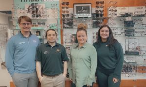
Dr. Hitchmoth with The RETeval device from LKC Technologies. Dr. Hitchmoth says this device is changing how she cares for diabetic patients.
Sponsored Content
By Dorothy Hitchmoth, OD, FAAO, ABO, ABCMO Dipl.
April 26, 2023
Electroretinography (ERG) has long been considered a valuable tool in caring for patients who have diabetes.1–4 However, the move to portable, hand-held, quicker ERGs has made this valuable diagnostic and monitoring tool a practical staple in a growing number of optometric practices.
What’s more, this technology has demonstrated tremendous benefits on two fronts: it’s a clinical powerhouse and reimbursement for the most commonly administered test is a reliable revenue stream for those of us working to save vision loss through early disease detection.
The most commonly used procedure code comes with an average payment of $128, and the diagnostic code choices number more than 560. These broad ICD-10 code set options help clinicians create a progress note that demonstrates medical necessity.
Interestingly, as I’ve expanded the use of hand-held ERG in my own practice, I started reflecting on how we view coding overall—with any technology and in almost every disease state that walks through our doors. Too often, we begin with a diagnosis and work backwards, which is counterintuitive to why we need advanced testing to begin with. There are numerous visual complaints and other symptoms, as well as diagnoses, that should drive testing.
Completing a proper medical history is paramount in the justification to utilize additional tests, such as ERG, for early detection and differentiation of either symptoms or diagnosis. ERG testing offers a striking example of how simple it can be to approach a patient with a “codable” concern or diagnosis by either symptom or a structurally identified disease state.
Clinical Benefits
Electroretinography objectively evaluates the functional abnormalities of the retina, while structural imaging reveals the anatomy of the retinal tissue. While both functional and structural assessments have their benefits, functional changes generally appear well before structural changes5-7 and are often accompanied by symptoms such as blur, glare, photopsia and scintillations. In studies comparing the ability of ERG and structural imaging to evaluate sight-threatening diabetic retinopathy (DR), ERG outperformed traditional imaging by predicting which patients would likely need subsequent medical intervention.5-7 So, clarifying visual function during the medical history is just as important as examining the retina in these patients.3,4
Glaucoma Parallels
The way many optometrists approach glaucoma is an example of how we should approach DR. There is controversy and payer variation with regard to coding in glaucoma suspects, particularly as it relates to the medical necessity to run an OCT based on a patient being a glaucoma suspect. However, OCT measure has become a standard in the management of glaucoma. This is because it is congruent with our belief that the patient may have glaucoma based on other clinical findings.
For example, a patient may have a retinal nerve fiber layer defect, drance hemorrhage, visual field defect or family history. If so, we code for these corollary findings in order to differentiate from other conditions and to follow for change over time. Ultimately, we get the answer we seek regarding the patient’s glaucoma status, but a variety of tests help us determine a clinical treatment plan.
The same philosophy applies to your approach in the care of people with diabetes. Consider the countless data points that we encounter in patients who have diabetes, and remember, diabetic eye disease runs along a spectrum and the earliest findings are often functional, so having the tools to help you detect abnormal retinal activity can even be considered an imperative in the practice of optometry. —Dorothy Hitchmoth, OD, FAAO, ABO, ABCMO Dipl.
The RETeval ERG device by LKC Technologies is the only FDA-cleared, portable, battery-operated, non-mydriatic ERG testing instrument on the market in the U.S. It has skin rather than corneal electrodes, adjusts for pupil size in real time and doesn’t require dilation. In my opinion, it is one of the easiest-to-use instruments in the optometrist’s toolkit because it’s portable, fast, tech-friendly and enhances confidence when caring for patients who have diabetes and are at-risk for DR.
The RETeval device offers an objective assessment protocol for measuring likely DR development and progression risk. By combining a retinal cell stress measure and a pupil light response, the device allows me to rapidly and noninvasively assess patients at risk for DR, as well as disease progression, in those with readily identifiable structural changes in the retina. Keep in mind, early visual or functional losses noted during the medical history warrants just as much investigation as the finding of an intra-retinal hemorrhage. 4 Importantly, interpreting results is easy—a score of 23.4 or higher indicates an 11-fold increase in the likelihood that the patient will require medical intervention within three years.6
Practice Management Benefits
As with any technology, the RETeval requires an investment, but comparatively speaking, this technology is exceedingly affordable and, with a fair reimbursement, it’s one of the most finance-friendly acquisitions I’ve made in my practice. A technician can perform a RETeval exam in both eyes within minutes,6 making it one of the most efficient tools available in optometry.4 Plus, it’s completely objective and doesn’t cause patient frustration. Patients tolerate the test and love the fact that they do not have to respond or provide the “right answer.” Many of the barriers in other objective tests are removed to include patients with language and educational barriers, as well as those who fatigue, or have difficulty staying in a chin rest or moving from room to room. The RETeval device can literally fit in your pocket and be accessed almost as easily as an ophthalmoscope.
Clinical Coding Approach
The most commonly used CPT code is 92273, electroretinography (ERG) with interpretation and report; and includes ffERG, Flash ERG and Ganzfield ERG.8 It is important to note that Medicare does not consider this test mutually exclusive to other diagnostic tests. This means that you can perform ERG, visual fields and other photo-imaging on the same day, which leads to a more comprehensive, accurate investigation in fewer visits. Fewer visits at a higher revenue per encounter results in improved margins to the practice while lowering the healthcare burden to the system and patient overall. The use of ERG might even be considered a best practice in this regard.
In talking to my colleagues, one thing I’ve noticed is that, when coding, practices often start with a disease state and work backwards. This approach to clinical care is reactive as opposed to proactive. This inspired me to partner with LKC to develop a billing and coding guide that encourages clinically-minded coding that’s driven by unanswered concerns and investigating symptoms that walk through our door, rather than by estimating what disease the patient may develop. This may sound esoteric, but flipping how we think about retinal disease is actually not that complicated, and is a much more common-sense approach to patient care, practice management and medical chart audit worries.
Essentially, every encounter we have with patients is an investigation of guilty until proven innocent when complaints are present. For example, in the case of ERG, ask yourself, “What do I see or hear that concerns me, and would a functional test that detects retinal dysfunction help me uncover either early-stage disease or progression?” If the answer is yes, then you have an opportunity to treat the patient with intent and precision. For patients with diabetes who have glare or photopsia and an abnormal or asymmetric ERG, we can further counsel about the importance of glycemic control and help them understand that their eye disease is often invisible in early stages.
You can also take a similar approach with patients who have evident diabetic retinopathy. You can use ERG to predict who is more likely to progress so you can tailor visit frequency and ensure that you will recommend the right time to refer to a surgical retinologist. This is becoming increasingly important because earlier anti-VEGF treatment is emerging in the literature as a potentially sight-saving option. 9
There are 560 ICD-10 codes to choose from, but our simple coding guide will help make chair-side investigations simple. This shift in thinking could make a big difference as we deliver care in the face of the costliest epidemic of our time—diabetes.10
Prioritize Patient Care
ECPs always look for classic signs of diabetic retinopathy such as intra-retinal hemorrhages and exudates. However, it is important to remember that classification guides that have been used for years include findings such as venous beading, IRMA, microaneurysms and cotton wool spots. Even a single observation of one of these findings falls under the EDTRS definitions and would justify functional testing such as ERG.11 Moreover, artificial intelligence algorithms (AIA) are right around the corner and can identify early changes with remarkable accuracy. 12 But, adding ERG to your practice will help your practice continue to differentiate technologically. Notably, screening AIA does not detect early functional changes in the retina thus far and it doesn’t converse with patients. With this in mind, remember to listen to your patients (e.g., vision changes, blur, glare, photopsia) and observe them carefully for very early retinal changes. This simple approach will help you identify disease early, which ultimately can help you help your patient avoid the path of vision loss.
References
- Kim M, Kim RY, Park W, Park YG, Kim IB, Park YH. Electroretinography and retinal microvascular changes in type 2 diabetes. Acta Ophthalmol. 2020;98(7):e807-e813. doi:10.1111/AOS.14421
- Miranda M, Sánchez-Villarejo MV, Álvarez-Nölting R, et al. Electroretinogram Alterations in Diabetes? Electroretinograms. Published online August 9, 2011. doi:10.5772/23490
- Yonemura D, Aoki T, Tsuzuki K. Electroretinogram in Diabetic Retinopathy. Archives of Ophthalmology. 1962;68(1):19-24. doi:10.1001/ARCHOPHT.1962.00960030023005
- McAnany JJ, Persidina OS, Park JC. Clinical electroretinography in diabetic retinopathy: a review. Surv Ophthalmol. 2022;67(3):712-722. doi:10.1016/J.SURVOPHTHAL.2021.08.011
- Zeng, Y. et al. (2019). Br. J. Ophthalmol. 103, 1747–1752.
- Brigell, M.G., Chiang, B., Maa, A.Y. and Davis, C.Q. (2020). Translational vision science & technol¬ogy, 9(9), 40-40.
- Al-Otaibi, H., Al-Otaibi, M. D., Khandekar, R., Souru, C., Al-Abdullah, A. A., Al-Dhibi, H., … & Kozak, I. (2017). Translational Vision Science & Technology, 6(3), 3-3.
- Billing and Coding: Electroretinography (ERG) (A57677). Accessed April 1, 2023. https://www.cms.gov/medicare-coverage-database/view/article.aspx?articleId=57677&ver=5
- Nanegrungsunk O, Bressler NM. Prevention of vision-threatening complications in diabetic retinopathy: Two perspectives based on results from the DRCR Retina Network Protocol W and the Regeneron-sponsored PANORAMA. Curr Opin Ophthalmol. 2021;32(6):590-598. doi:10.1097/ICU.0000000000000799
- National Diabetes Statistics Report | Diabetes | CDC. Accessed April 1, 2023. https://www.cdc.gov/diabetes/data/statistics-report/index.html
- Wilkinson CP, Ferris FL, Klein RE, et al. Proposed International Clinical Diabetic Retinopathy and Diabetic Macular Edema Disease Severity Scales. Ophthalmology. 2003;110:1677-1682. doi:10.1016/S0161-6420(03)00475-5
- Lim JI, Regillo CD, Sadda SR, et al. Artificial Intelligence Detection of Diabetic Retinopathy. Ophthalmology Science. 2023;3(1):100228. doi:10.1016/j.xops.2022.100228
Dorothy Hitchmoth, OD, FAAO, ABO, ABCMO Dipl., is the owner of Dr. Dorothy L. Hitchmoth, PLLC in New London, N.H. To contact her: drdorothy@hitchmotheyecare.com


