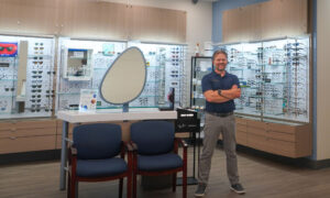
 By the NECO Innovation Center
Beneath the purview of Gary Y. Chu, OD, MPH, FAAO, Vice-President, Professional Affairs
March 29, 2023
epipole founder Craig Robertson, PhD, an expert in mathematics, computer science and artificial intelligence, created epipole in 2011 after working with Optos. He was predisposed to working in the vision space because he witnessed first-hand his grandmother’s rapidly deteriorating sight from diabetes. This experience never left him and motivated him to think outside the box to re-imagine the development of products that will aid in diagnosing retinal diseases and make a social impact with the company’s mission being the eradication of preventable blindness.
After many iterations and designs, the first camera was conceived after breakthroughs in solving how light interacts with biological tissue and the conundrum of understanding how to resolve the illumination and the optical axis on top of one another.
The team of engineers used early assessments from international trips2 and local eyecare practitioners3 to continuously improve the epiCam. The team valued the importance of testing their design in relation to resolution, performance, usability, comfort and ergonomics. This led them to develop a proprietary model eye to test their design and aid in training. This spirit of “innovate-test-improve” continues to be an area of focus today.
Overview of the Device
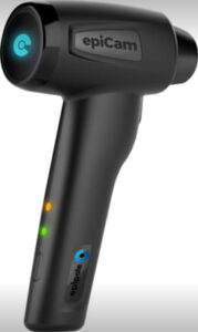
In essence, the epiCam, designed for the U.S. market is an ophthalmoscope that records continuous retinal images of the fundus and subsequently allows the eyecare practitioner to select the appropriate images to be used for diagnostic assessment. The camera is lightweight and ergonomically designed to fit a provider’s hand well. The examiner can change illuminants via a trigger system to toggle between normal non-coherent light, amber (589 nm) and deep red (660 nm). The amber light allows the practitioner to optimally view retinal blood flow and the deep red aids in enhancing the retinal choroidal layer.
The epiCam is connected wirelessly to a computer via a WiFi system to ensure the high-speed capture and transfer of the fundoscopic images. The retinal imagery can be viewed in real-time on the epiCam or the computer. Similar to other ophthalmic imaging devices, patients are registered to the system to ensure recorded retinal videos are assigned to the appropriate patient for review on the computer. When reviewing the retinal videos, specific images can be captured for your report.
Science of the Device
The quest for providing detailed imaging of the fundus of the eye is driven by the need to facilitate, wherever possible, differential diagnosis of various retinal pathologies. In the past, fundus imaging has been dominated by ophthalmoscope-type devices that operate in a mode known as fundus photography. Scanning laser-type light sources can be used in these types to acquire individual, select retinal images and video4.
For example, the video image can help document:
- blood transport through pulsing and distortion of blood vessels.
- change in deformation of the optic nerve head and microscopic movements in nerve tissue.
- monitoring and estimate of the intracranial pressure (ICP) for neurodegenerative diseases such as Parkinson’s and Alzheimer’s.5
The reason that, so far, there has not been any known or commercially successful retinal ‘video recorder’ is none other than the fact that the acquisition of video imaging of the retina is faced with certain challenges. These include technological and optical-design challenges, which include the following:
- Resolution and overall image quality for digital video imaging have always lagged behind still imaging
- Throughput of digital video imaging places significantly greater demands on the device’s hardware, storage and connectivity with existing IT infrastructure
- A focusing mechanism that needs to address the effects of variable refractive error of the subject’s eye
- An optical delivery of non-coherent radiation that can achieve lateral resolution down to a few microns
- Counteracting the mismatch effects caused by the natural movement of the imaging apparatus with respect to the subject eye
This device implements an optical design based on a relay of forward-scanning optics delivering non-coherent deep red light on the retina. It incorporates advanced sensor technologies to aid in focusing through the optics of the eye. Lenses and light sources use lightweight materials, and deliver a handheld, battery-powered and reliably interconnected device.
What Do ODs Think?
Feedback from optometrists and optometry students extolled the ergonomics and comfort of the epiCam in the examiner’s hand and the continuous video capacity, which ensures nothing is missed while examining a patient. The instrument is not meant for all practices or all patients.
It is much appreciated when examining patients with cognitive impairment, individuals with trouble maintaining fixation and patients with limited mobility, including supine patients, as well as having in-clinic applications.
The portability of the epiCam is its greatest asset, along with the flexibility afforded by the multi-illuminant operation to help assess the subtleties of fundus images that are captured. It also allows creativity in how an optometrist can utilize the instrument, especially in nursing homes, care for special populations, home-care practices and medical mission trips. Many commented that it is very similar to using an ophthalmoscope and has enhanced care in such settings.
Characteristics of the EpiCam
- Continuous live, real-time video capturing of posterior pole
- Ability to capture images during an examination or when viewing the video of the retinal examination
- Amber light (589 nm), Deep Red light (non-IR, 660 nm)
- Portable and handheld
- Lightweight
- Designed with ergonomics and comfort in mind
- Durable, rechargeable
- Can be used in lighted rooms
- Affordable
- FDA approved
- Can be used undilated
- 45 degree view
- Handheld design allows for straightforward panning and tilting for a broader field of view to help locate areas of interest
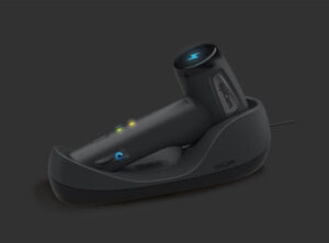
There is a learning curve, but it is not insurmountable. The epiCam’s cradle ensures the device is always charged and the assessment of images is quickly and easily viewed. However, there is currently no integration into an electronic health record or image management system.
Final Thoughts
If the pandemic has taught us anything, it is that we need to think outside the box, especially in a world where handheld technologies are rapidly outpacing desktop imaging device adoption. epipole has delivered a fascinating and innovative device, and we look forward to seeing more from them in the future.
References
1. Robertson C, Good Business, retrieved from https://theophthalmologist.com/subspecialties/good-business, Accessed 3/8/2023.
2. Using a new low-cost retinal camera to screen for diabetic retinopathy in rural eye camps – L Nozad, S Ruvuma, C Davey, A Blaikie; Presented at Pantheo Eye Center Annual Congress, 2019 (winner of the Alistair Fielder Prize)
3. Remote Interpretation of Optic Nerve Head Cupping from Images Acquired Using A New Low-Cost Retinal Camera (epiCam) Capable of Being Powered and Operated from an Android Based Mobile Phone – S Watson, F Burgess, A Blaikie; presented at Scottish Ophthalmological Club, September 2016
4. epipole Patent: Image Acquisition Apparatus, https://worldwide.espacenet.com/patent/search?q=pn%3DEP2911573A1, Accessed 3/8/2023