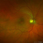
Wide-field retinal imaging photo, courtesy of Optomap. Dr. Murphy says that the technology can be a great tool in diagnosing patients, but that dilation still is essential.
By Cheryl G. Murphy, OD
April 13, 2022
When Review of Optometric Business asked me to write an update on my May 3, 2018, article, “Can Advanced Technology Replace Dilation?,” which was one of the most viewed articles on the site, I questioned whether a rewrite was necessary. I reread the article and still agreed with much of what I wrote. However, I do have a couple key points to add.
An Advantage: Pandemic Protection
Over the last four years, and particularly during the pandemic, I developed a deeper appreciation for wide-field digital imaging. Before it was understood how COVID-19 was transmitted, wide-field digital imaging via the Optomap helped me monitor the eye health of my patients while putting them and myself at lower risk for infection. This was possible because the private practice where I work made the decision to perform an Optomap on every patient out of courtesy (free of charge) during the worst of the pandemic.
The reasoning was that we would use it to minimize the amount of time doctors spent in close range of each patient. During this period of complimentary Optomap, patients who may not have agreed to Optomap in the past (perhaps because of the $45 out-of-pocket fee) got a chance to try it out and “take a tour” of the inside of their eye for the first time. Many were fascinated with how much we could see inside the eye. It was a great conversation-starter, invaluable to patient education, and helped us to capture baseline images on patients who may not have had them otherwise. I started to enjoy reviewing Optomap images, and found myself disappointed once the fee was reinstated and not everyone opted for imaging the following year.
A Disadvantage: What Optomap Can Miss
As my usage of Optomap grew, so did my false sense of security that if the Optomap looked good then everything must be alright and the patient’s eyes must be healthy. As I noted in my previous article, in the past I saw firsthand how wide-field images can miss retinal tears. However, recently, I had a different type of scare involving the anterior segment.
I saw a family friend for an exam. When I see friends and family, I tend to catch up first and then do a thorough exam, so we chatted for a few minutes and then I got started. I did the refraction, reviewed their Optomap images with them, tested their pressures using Goldmann, checked for dry eye since the fluress was in there anyway and then I did one final sweep of the anterior segment using the slit lamp. It was on the temporal iris of the left eye, the last place I was going to look before concluding the exam, where I saw something strange: a non-pigmented elevation on the iris with blood vessels inside it.
It was not like anything I had seen in my 18 years of practicing. I examined the patient four years ago, and they had gone to another friend who is an OD two years before this exam, so I contacted the other OD to see if they had seen it or anything else wrong with the left iris two years ago. They assured me they had not, which led me to believe this was new, and therefore, growing at a relatively rapid rate. I snapped a quick photo holding my iPhone up to one ocular of the slit lamp and performed an anterior segment OCT on the iris and angle to see the depth of the elevation and compare the thickness of the temporal iris in the left eye to the nasal iris of the same eye. It was definitely thicker and an elevation.
Long story short, after two referrals and a biopsy by an ocular oncologist, it was determined to be a malignant iris melanoma. A full body PET scan revealed the iris melanoma was the primary site of growth, and it appears it has not metastasized from or to anywhere else. The ocular oncologist is performing radioactive plaque therapy on the patient next month.
Had I not been so diligent about doing a thorough exam, including a careful evaluation of the anterior segment under magnification using biomicroscopy, I might not have saved this asymptomatic patient’s life since the growth was amelanotic (non-pigmented) and blended in with the patient’s blue iris. It could only be seen with the magnification, not with the naked eye.
Beware of Making Snap Judgements
My point is, the Optomap images looked good, so I almost jumped to the assumption that the patient’s eye health was normal when it was not. Good thing I was diligent and checked! This may seem obvious, but I bet I am not the only one who has been tempted to assume that all is well with an asymptomatic patient who corrects to 20/20 easily and whose Optomaps look normal. Technology doesn’t replace being a good doctor, it just aids a good doctor in doing an even better job.
Other Articles to Explore
In conclusion, while I love Optomap for many reasons, it is important to realize it is not the end-all-be-all and it does not take the place of us doing a thorough eye exam of both anterior and posterior segments. Don’t put blinders on to other aspects of the eye just because the Optomap looked great. I find most optometrists are good doctors who truly care about their patients and do the right thing. However, it’s important to remember that advances in technology should enhance and add to our already-great eye exam and not replace parts of it. As awesome as Optomap is, there are limitations to the technology, and that is one of the reasons it is supplemental to, and not a substitute, for a full, comprehensive eye exam with dilation.
 Cheryl G. Murphy, OD, practices in Lake Ronkonkoma, N.Y. You can like her on Facebook or follow her on Twitter @murphyod. To contact her: murphyc2020@gmail.com.