By Cheryl G. Murphy, OD
May 9, 2018
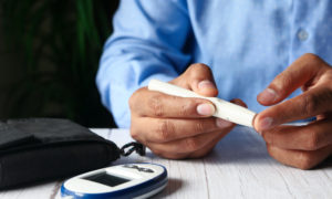
Advanced technology, like Optomap, offers greater power to diagnose and manage ocular disease, but think twice before you use it as a replacement for dilation. There is no technology that replaces dilation for thoroughly assessing eye health, and protecting your practice in the event of a lawsuit for a missed diagnosis that leads to vision loss.
The other day, I passed by a well-established optometry practice. This place is highly regarded, and I am sure that regard is well deserved. However, I was struck by a new sign in the window: “No Dilation Necessary. New State of the Art Technology Available.” Doubtless, “no dilation” appeals to patients who don’t like getting dilated, with the time and discomfort required. But there are dangers to this approach.
I assume they are talking about Optomap. According to its web site, Optomap is “a unique technology that captures more than 80 percent of your retina in one panoramic image.” The site also states that it “gives a ultra-widefield retinal image while traditional imaging methods typically only show 15 percent of [a patient’s] retina at one time.”
I work at a practice that has Optomap. I think it is an extremely valuable screening tool, but I am careful in how I explain it when offering it to patients. I like saying, “It is an alternative to, but not a substitution, for a dilated eye exam.”
I say that it is not a substitution for a dilated eye exam because, as anyone who uses Optomap has experienced, there are limitations to the device. I think one of the main limitations is that it does not image all of the retina. There are parts of the retina superiorly and inferiorly that are missed by Optomap, usually because those areas seem to be limited or blocked by the upper and lower lids.
I have personally seen this become a problem. I had a patient with a history of retinal tears, and retinal surgery to repair those tears. The patient wanted to be dilated, but I also thought it would be a good idea to do Optomap to photodocument much of the retina (a great advantage that Optomap gives us.)
After reviewing the Optomap images, it looked like the old areas of retinal tears previously repaired by surgery were stable, and had not changed or expanded in any way. However, I then looked in with BIO and a Panretinal 2.2 lens (as opposed to the more traditionally used 20D fundus lens), and I noted fresh areas of retina changes that looked problematic in the areas that Optomap didn’t image (because they were blocked the lids.) I sent the patient back to his retinal specialist, and they confirmed that these were new areas (that they also had not previously documented), and secured those areas with laser treatments.
Supporting my personal experience, in 2017 a study was published in Seminars in Ophthalmology, which examined 36 eyes of 34 patients, and retrospectively compared the use of Optomap to indirect ophthalmoscopy in evaluating patients with a history of with non-traumatic rhegmatogenous retinal detachment. The study found that “ultra-widefield imaging failed to detect retinal holes in the superior and inferior quadrants in 11.1% and 19.4% of cases, respectively [and in] postoperative imaging, UWF photos did not detect retinopexy which was ophthalmoscopy-visible both superiorly and inferiorly in 19.4% of cases.”
The study concluded that while “ultra-widefield imaging is a useful adjunct for documentation of rhegmatogenous retinal detachments and their postoperative repair, the detection of retinal holes, tears, and postoperative scarring is poor, especially in the inferior and superior periphery.”
Had I not seen for myself what Optomap can miss, I would not be so quick to judge a sign outside of a practitioner’s office saying, “No dilation necessary.” I do think that Optomap is an amazing tool that brings eyecare professionals a tremendous advantage in that we can finally screen better for eye conditions and illnesses, particularly in those patients who, year after year, come up with some excuse as to why they can’t be dilated the day of their exam and then disappear, not returning on another day with a driver as they promised.
Optomap is also a chance for us to gain more revenue for the practice since Optomap often is a separate, out-of-pocket charge that insurance doesn’t cover, from between $35 and $50. My only qualm is the wording that we as practitioners use, and the way in which we present Optomap. I think we have to be careful to not say it is a substitution for dilation.
Patients (or people driving by) may easily misinterpret our words, or a sign outside of an office, saying that dilation is not necessary. I think we can all agree that dilation is still necessary in many cases, like those of patients with a previous history of retinal thinning, holes, tears, detachments or surgeries. Let’s not let new technology deter us too far away from the more tried, true and thorough way of examining eyes. Instead, let it complement the more traditional method of indirect ophthalmoscopy, and act as a way for us to photodocument as much as we can.
How has Optomap helped your practice grow? Has it enhanced patient education, photodocumentation and boosted revenue? Have you found any limitations personally with the device? Do you believe the technology will advance to allow us to truly photodocument the entire inside of the eye?
 Cheryl G. Murphy, OD, practices in Glen Cove, Syosset and Lake Ronkonkoma, N.Y. You can like her on Facebook or follow her on Twitter @murphyod. To contact her: murphyc2020@gmail.com.